 Brain is the anterior end of spinal cord that has enlarged to take care of the sense organs which are located on the anterior end of the body in a bilaterally symmetrical animal that moves ahead in an anterior-posterior axis. This is called cephalon, which as it evolves further, divides into three parts, namely, prosencephalon, mesencephalon and rhombencephalon. As the brain develops further by increasing the number of neurons, it further divides into different parts, each one having assigned its own specific function.
Prosencephalon divides into Telencephalon and Diencephalon, the former includes olfactory lobes (Rhinencephalon) and Cerebral hemispheres that coordinate the activities of the entire brain. The roof of cerebrum is called Pallium and the floor that generally contains nerve fibres is known as corpusstriatum.
Diencephalon is a small part of brain, generally covered by enormously enlarged cerebral hemispheres. This is an extremely important part of brain which functions as switch board to cerebrum. Dorsal part of diencephalon is called epithalamus and the ventral part hypothalamus while the lateral parts are called thalami that contain relay centres to connect dorsal and ventral parts of thalamus.
Anterior part of epithalamus contains a glandular area called anterior choroid plexus(Tela Choroidea) which secretes cerebro-spinal fluid. Two dorsal processes of epithalamus, the anterior paraphysis supports parietal body and the posterior epiphysis bears pineal body. These two bodies function as photoreceptors in lower vertebrates and gradually transform into endocrine organs and biological clock in higher vertebrates.
The ventral hypothalamus has the optic chiasma (crossing of optic nerves) on the anterior side and a ventral median evagination called infundibulum which supports pituitary gland or hypophysis. There is an olfactory area, mammillarybody on the posterior side of hypothalamus.
Mesencephalon is concerned with sight and hearing. Its dorsal side is called Tectum and the ventral fibre bundles are called Cruracerebri or cerebral peduncle. The tectum has a pair of bulging optic lobes on the anterior side and a pair of auditory lobes on the posterior side. In lower animals auditory lobes are insignificant and optic lobes are prominent. This is called Corporabigemina. Higher vertebrates such as mammals and snakes have corpora quadrigemina, which means they have optic and auditory lobes of equal size.
Metencephalon is called cerebellum which is quite enlarged in active animals. In mammals cerebellum contains bundles of branching fibres of white matter called ArborVitae. The bulging ventral side of cerebellum is called pons varolli and it contains criss-crossing fibres of neurons.
Myelencephalon or medullaoblongata is the posterior part of the brain which does not undergo much modification in vertebrates since it controls the autonomic functions of body. The ventral side contains RAS (Reticular Activating and Inhibiting System) which keep the brain attentive and awake. Dorsal side exhibits the posterior choroid plexus, which secretes cerebrospinal fluid that flows into the brain ventricles and to meninges through a median Foramen of Megendie and the paired Foramina of Luschka. Medulla is attached with cranial nerves which bring sensory impulses from the body.
Brain is hollow inside; the cavities are called ventricles which are lined by ciliated epithelium, ependyma. Ventricles of the two cerebral hemispheres are called lateral ventricles, or Telocoel or I and II ventricles which are connected together with a foramen of Monro. The third ventricle extends from diencephalon to mesencephalon and the IV ventricle is larger inside metencephalon and myelencephalon. The third and fourth ventricles are connected together by a tube-like connection called Iter or aqueduct of Sylvius.
MENINGES
Meninges are protective layers around the brain. The outermost layer is fibrous duramater (meaning tough mother) which, though tightly attached to the periostial layer of skull, still encloses a narrow epidural space that is filled with perimeningeal fluid. The second layer under dura mater is Arachoid, so named because of spider web like appearance due to presence of villi for the absorption of cerebrospinal fluid. Between the dura mater and arachnoid exists the subdural space and between arachnoid and the lower pia mater is the subarachnoid space. The innermost layer of meninges is the delicate pia mater which is intimately attached with the brain tissue and extends deep into the sulci and fissures. It carries blood vessels and nerves.
The three separate meningeal layers are found in mammals only, while in amphibia, reptiles and birds, arachnoid and pia mater fuse to form a single pia-arachnoid layer below the subdural space. Fishes have a single meninx primitiva that is separated from the skull bone by perimeningeal tissue.
CYCLOSTOME BRAIN
Cyclostome brain is very primitive owing to their parasitic and detritus feeding habits. Cerebral hemispheres are small and smooth. Olfactory lobes are well developed as these animals detect suitability of their hosts by acute sense of smell. For the same reason thalamus, which contains olfactory relay centres, is enlarged with a prominent median olfactory area called habenula. Optic lobes are small because of primitive or rudimentary eyes. Cerebellus which is related with balance and posture is reduced due to parasitic mode of life. Medulla oblongata is quite well developed and receives six pairs of cranial nerves but there is no pons varolli on the ventral side. Pineal and parietal bodies are present in lampreys but absent in hag fishes.
FISH BRAIN
Active bony fishes and sharks have well developed brain but bottom dwelling fishes have reduced brain organs. Olfactory lobes are large in sharks and they can detect their injured prey by the smell of blood from a distance of about a kilometer. But in majority of bony fishes optic lobes are reduced. Cerebral hemispheres are quite large but smooth and white. Pineal and parietal bodies are generally reduced in fishes. On the ventral side of diencephalon, there is saccusvaculosus posterior to the pituitary that serves as sense organ.
Optic lobes are well developed as most fishes are gifted with large eyes but in deep sea fishes they are reduced. Cerebellum is highly enlarged in sharks as well as in active bony fishes and also has lateral extensions called restiformbodies or auricularlobes which connect medulla with cerebellum. They help in maintenance of balance. Cerebellum is smaller in rays, lung fishes, ganoid fishes and deep sea fishes. Medulla oblongata has no particular variation except in deep sea fishes in which there are large vagal lobes on the lateral side which receive impulses from taste buds that are scattered all over the body as pit organs.
URODELE BRAIN
Urodele brain is primitive and reflects their sluggish nature and under-developed sense organs. Olfactory lobes, optic lobes and cerebellum are reduced and cerebral hemispheres are also small and smooth. Pineal and parietal bodies are present but reduced. There is no saccus vaculosus and corpus striatum is weak.
ANURAN BRAIN
Frogs and toads possess a better developed brain as compared with urodeles. Olfactory lobes are large and fused at base that gives better sense of smell to frog. Cerebral hemispheres are larger with a centralized gray area called Archipallium that controls olfactory sense, while the lateral areas are white and are called paleopallium. Parietal body is reduced but pineal is well developed and probably a photoreceptor. Ancient amphibians possessed a third eye over the pineal-parietal complex. Optic lobes are well developed but there are no auditory lobes and hence the mesencephalon is called corpora bigemina. Cerebellum is reduced but medulla is enlarged to make the brain a reflex brain or spinal brain.
REPTILIAN BRAIN
Brain becomes large by the enlargement of corpus striatum. Cerebral hemispheres are large and oval but the surface is white and smooth except in crocodiles which develop gray matter called neopallium similar to mammals. Olfactory lobes are well developed in snakes and lizards which have olfactory sense organs on the tongue, but reduced in turtles and crocodiles. Parietal body is well developed in lizards and in Sphenodon it lies under a lens-like transparent area called the third eye. Pineal body is absent in crocodiles. Optic lobes are well developed in all reptiles and corporaquadrigemina is found only in snakes. Cerebellum is reduced in all reptiles owing to their creeping habit.
BIRD BRAIN
Bird brain is characterized by enormous enlargement of cerebral hemispheres, optic lobes and cerebellum. Cerebral hemispheres become enlarged owing to enlargement of corpus striatum which is called hyperstriatum but pallium is thin and surface has only white matter. Olfactory lobes are highly reduced, attached to the anterior end of the cerebral hemispheres. Optic lobes are enormous as birds are gifted with the best eye sight in animal kingdom. Parietal body is absent and pineal small in most of the birds. Being bipedal and flying animals, birds need good control over muscles and tendons, which comes from a trilobed highly enlarged cerebellum. The middle lobe of cerebellum is called vermis as it has transverse folds and the lateral lobes are called flocculi. Birds’ brain is instinctive brain that can carry out complex but stereotype functions such as nest building. Spinal cord still has equal control over the body.
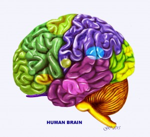
MAMMALIAN BRAIN

Mammalian brain is highly developed and keeps complete control over the body functions. Cerebral hemispheres are enormously enlarged and the surface is folded into depressions (sulci) and raised portions (gyri) so that large surface area can be accommodated in the small space of the skull. Gray matter has spread to the surface which is called neopallium. Two cerebral hemispheres are connected by thick bundles of nerve fibres called corpus callosum, which is not found in monotremes and marsupials. Olfactory lobes are highly enlarged, so that mammalian brain is sometimes called nose-brain. Parietal body is absent and pineal is usually present except in animals like Armadillo, Sirenia and Edentates. Mammals being active animals, cerebellum is highly enlarged and trilobed and all lobes possess gyri and sulci. Nerve cells form bundles of branching fibres called ArborVitae. Medulla is short as compared to the large brain but ponsvarolli is enlarged.